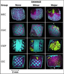
 From Cochrane et al. Electron microscopy images of bracket pads. Green is calcium, purple is silicon, orange is phosphorus. From Cochrane et al. Electron microscopy images of bracket pads. Green is calcium, purple is silicon, orange is phosphorus. BY WILLIAM R. PROFFIT, DDS, PHD STUDY SYNOPSIS This study at the University of Melbourne, Australia, provides new data for the prevalence of enamel damage from debonding brackets, and relates such damage to the type of bracket, the type of adhesive, and the surface preparation of the enamel for bonding. Four groups of brackets and bonding techniques were examined. All brackets were GAC Innovation – either the metal “R” version or the ceramic “C” version. All brackets were bonded and removed in the setting of 5 private orthodontic practices. 437 total brackets from anterior teeth were analyzed. The four in-vivo bracket and bonding protocol combinations were:
Debonding was accomplished for metal brackets with a debonding instrument (444-761 bracket lifter from Unitek) and for ceramic brackets with a debonding plier. Only maxillary canine to canine brackets were collected. The back of each bracket was visualized with scanning electron microscopy at 60 x magnification. An elemental map was made using dispersive x-ray spectrometry to detect calcium, phosphorus, aluminum, and silicon. Calcium and Phosphorus together indicated the presence of enamel that had sheared from the tooth. Areas of bonding material and enamel were mapped, the amount of bonding material was categorized, and bracket fracture was tabulated. The results:
WHAT THE PROFESSOR THINKS
This is a particularly interesting study because it analyzes data from patients treated across several orthodontics clinics, rather than just from laboratory testing. Despite some limitations, clinical orthodontists can use the following points to help inform their practice:
Article Reviewed: Cochrane NJ, Lo TWG, Adams GG, Schneider PM. Quantitative analysis of enamel on debonded orthodontic brackets. Am J Orthod Dentofac Orthop 152:312-319, 2017.
2 Comments
|
Curated by:
Tate H. Jackson, DDS, MS CategoriesArchives
October 2019
|
 RSS Feed